Technology

Ophthalmic Associates use the latest ophthalmic patient data management, imaging and laser technology to ensure patients have the most up to date case, such as:
- Zeiss Forum Software with retina and glaucoma workplaces
- Zeiss IOL Laser 700 optical biometer with the latest generation IOL calculation formulas and anterior and posterior corneal (Total Keratometry) measurements
The IOL Master is a modern, non-contact machine used for taking measurements of a patient’s eye in preparation for cataract surgery. These measurements are used to calculate the suitable type and strength of the new intraocular lens which will replace the natural lens during surgery. It is considered the “gold-standard” machine for pre-operational eye measurements. Every eye is unique, and it is important to obtain measurements as accurately as possible so that a patient can achieve the best possible outcome for their vision.
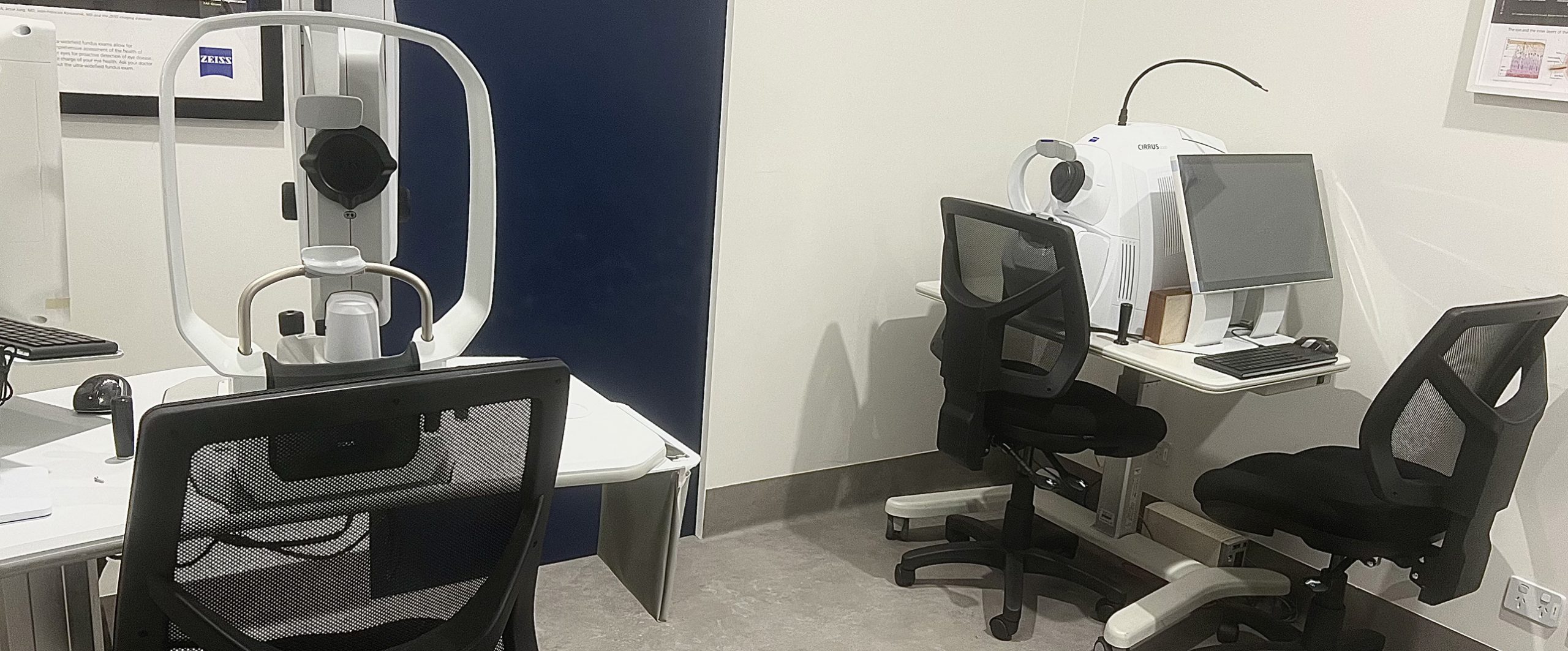
- ZEISS CIRRUS 6000 OCT spectral-domain OCT
The OCT machine enables a non-invasive examination of the posterior structures of the eye by using reflections of light to provide a cross-sectional image. It is a diagnostic device that aids in the detection and management of ocular diseases.
We use our OCT machine frequently to monitor macula or optic nerve disease in our patients. By comparing the structure and thickness of a patient’s scan against a normal, healthy eye, pathology can be detected, even at early stages.
- ZEISS CLARUS 700 ultra-wide field True Colour fundus imaging system (advanced retinal camera) with wide field fundus fluorescein angiography
This specialized camera takes highly detailed images of the back surface of the eye (the retina). This enables Eye Specialists to keep a record of the health of the eye as well as detect any small changes over time. The camera features an ultrawide lens that enables coverage over a wide area, as well as specialized filters that enable detection of different retinal layers.
- Zeiss VISUCAM 524 retinal camera with fluorescein angiography
Fundus Fluorescein Angiography is a diagnostic procedure, used to examine the blood vessels of the retina. Fundus angiography is often performed to confirm the initial diagnosis of a patient with retinal or macular disease.
- Zeiss Humphrey Field Analyzer 3 with SITA Faster visual field-testing algorithm and Guided Progression Analysis
The HVFA reveals the extent of a patient’s peripheral vision. It can expose early stages of disease and is used for the following:
- Diagnosis of an Ocular Disease and Glaucoma as this displays characteristic patterns of visual loss.
- Assessment of neurological disease-causing visual loss.
- Monitoring the progression or recovery of ocular and neurological disease.
- Drivers who are 75 years of age or older require a field test as part of their RTA form. The purpose of the field test is to rule out the presence of significant visual field loss, and therefore confirm whether they are fit to drive.
- Zeiss VISULAS green retinal photocoagulation Laser with both slit lams and indirect capabilities for treatment macula and peripheral retina
- Ellex Tango Reflex SLT/YAG Laser & Ellex UltraQ Reflex Yag Laser
- Slit-Lamp
The slit-lamp consists of an adjustable beam of bright light and a high magnification microscope for the examination of the anterior (front) and posterior (back) parts of the eye, as well as being able to measure the Intra-Ocular Pressure (IOP) of each eye. The Slit-Lamp is a vital tool that you will find amongst all ophthalmic practices.
Technology
Ophthalmic Associates use the latest ophthalmic patient data management, imaging and laser technology to ensure patients have the most up to date case, such as:
- Zeiss Forum Software with retina and glaucoma workplaces
- Zeiss IOL Laser 700 optical biometer with the latest generation IOL calculation formulas and anterior and posterior corneal (Total Keratometry) measurements
The IOL Master is a modern, non-contact machine used for taking measurements of a patient’s eye in preparation for cataract surgery. These measurements are used to calculate the suitable type and strength of the new intraocular lens which will replace the natural lens during surgery. It is considered the “gold-standard” machine for pre-operational eye measurements. Every eye is unique, and it is important to obtain measurements as accurately as possible so that a patient can achieve the best possible outcome for their vision.
- ZEISS CIRRUS 6000 OCT spectral-domain OCT
The OCT machine enables a non-invasive examination of the posterior structures of the eye by using reflections of light to provide a cross-sectional image. It is a diagnostic device that aids in the detection and management of ocular diseases.
We use our OCT machine frequently to monitor macula or optic nerve disease in our patients. By comparing the structure and thickness of a patient’s scan against a normal, healthy eye, pathology can be detected, even at early stages.
- ZEISS CLARUS 700 ultra-wide field True Colour fundus imaging system (advanced retinal camera) with wide field fundus fluorescein angiography
This specialized camera takes highly detailed images of the back surface of the eye (the retina). This enables Eye Specialists to keep a record of the health of the eye as well as detect any small changes over time. The camera features an ultrawide lens that enables coverage over a wide area, as well as specialized filters that enable detection of different retinal layers.
- Zeiss VISUCAM 524 retinal camera with fluorescein angiography
Fundus Fluorescein Angiography is a diagnostic procedure, used to examine the blood vessels of the retina. Fundus angiography is often performed to confirm the initial diagnosis of a patient with retinal or macular disease.
- Zeiss Humphrey Field Analyzer 3 with SITA Faster visual field-testing algorithm and Guided Progression Analysis
The HVFA reveals the extent of a patient’s peripheral vision. It can expose early stages of disease and is used for the following:
- Diagnosis of an Ocular Disease and Glaucoma as this displays characteristic patterns of visual loss.
- Assessment of neurological disease-causing visual loss.
- Monitoring the progression or recovery of ocular and neurological disease.
- Drivers who are 75 years of age or older require a field test as part of their RTA form. The purpose of the field test is to rule out the presence of significant visual field loss, and therefore confirm whether they are fit to drive.
- Zeiss VISULAS green retinal photocoagulation Laser with both slit lams and indirect capabilities for treatment macula and peripheral retina
- Ellex Tango Reflex SLT/YAG Laser & Ellex UltraQ Reflex Yag Laser
- Slit-Lamp
The slit-lamp consists of an adjustable beam of bright light and a high magnification microscope for the examination of the anterior (front) and posterior (back) parts of the eye, as well as being able to measure the Intra-Ocular Pressure (IOP) of each eye. The Slit-Lamp is a vital tool that you will find amongst all ophthalmic practices.

